Home

עדין תורם וידוי what is a fetal pole שפעת מכתש מרק

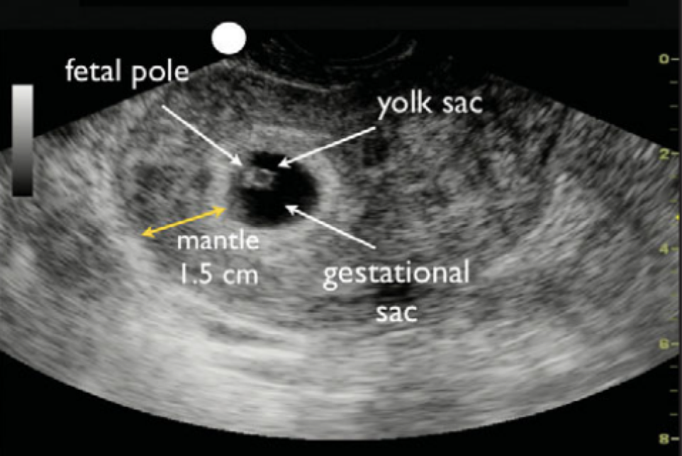
Derek Monette, MD MHPEd on Twitter: "IUP = yolk sac, fetal pole, or both.  #EMConf #FOAMed #FOAMus @Rachyhane @mghedus https://t.co/wrwQCF58JE" /  Twitter
Derek Monette, MD MHPEd on Twitter: "IUP = yolk sac, fetal pole, or both. #EMConf #FOAMed #FOAMus @Rachyhane @mghedus https://t.co/wrwQCF58JE" / Twitter

Figure, Normal gestational sac. Image courtesy S Bhimji MD] - StatPearls -  NCBI Bookshelf
Figure, Normal gestational sac. Image courtesy S Bhimji MD] - StatPearls - NCBI Bookshelf

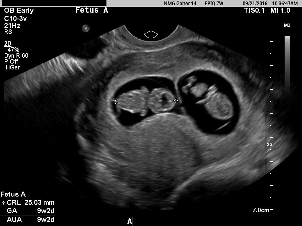
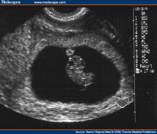
Fetal pole of intrauterine pregnancy, Twin A. | Download Scientific Diagram

Diagnostic Ultrasound in the First Trimester of Pregnancy | GLOWM

Diagnostic Ultrasound in the First Trimester of Pregnancy | GLOWM

Patience is key: Understanding the timing of early ultrasounds | Your  Pregnancy Matters | UT Southwestern Medical Center
Patience is key: Understanding the timing of early ultrasounds | Your Pregnancy Matters | UT Southwestern Medical Center

OBSTETRIC (First Trimester) | Department of Emergency Medicine | Saint John

EPOS™
EPOS™

Fetal Measurements
Fetal Measurements

Very large uterine fibroid in patient with an early intrauterine pregnancy  - Critical Care Sonography
Very large uterine fibroid in patient with an early intrauterine pregnancy - Critical Care Sonography

Ultrasound Case of the Month: Hydatidiform Mole — Brown Emergency Medicine

Figure 1. | Meningomyelocele: Early Detection Using 3-Dimensional  Ultrasound Imaging in the Family Medicine Center | American Board of Family  Medicine
Figure 1. | Meningomyelocele: Early Detection Using 3-Dimensional Ultrasound Imaging in the Family Medicine Center | American Board of Family Medicine

Ultrasound Video showing Gestational sac in the uterus without any fetal  pole or POC in it. - YouTube
Ultrasound Video showing Gestational sac in the uterus without any fetal pole or POC in it. - YouTube
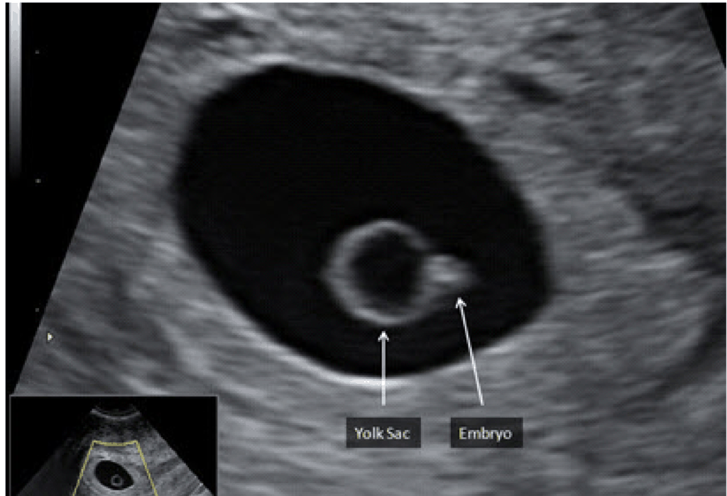
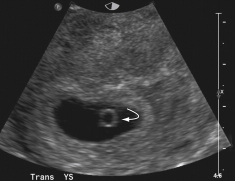
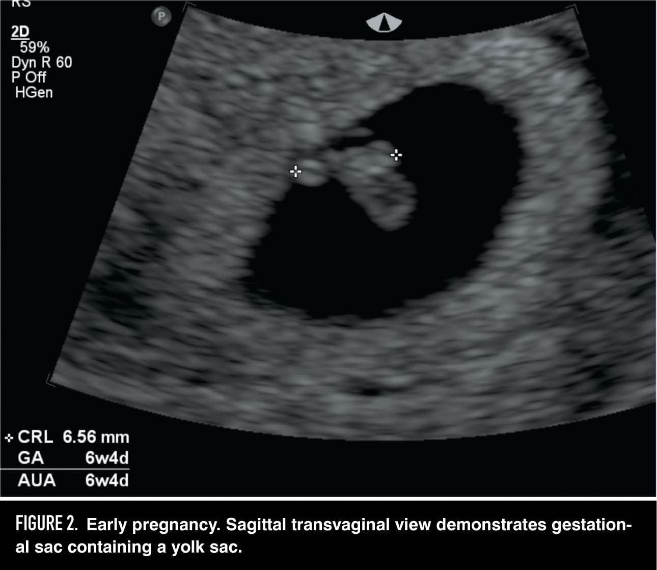
Trans-vaginal ultrasound image showing yolk sac and fetal pole. | Download  Scientific Diagram
Trans-vaginal ultrasound image showing yolk sac and fetal pole. | Download Scientific Diagram

Ultrasound of the Normal and Failed First-Trimester Pregnancy | Radiology  Key
Ultrasound of the Normal and Failed First-Trimester Pregnancy | Radiology Key

Case of the Month — Ectopic Pregnancy — Brown Emergency Medicine

When a pregnancy fails early

HCG Pregnancy Testing: Everything You Want To Know | Marie Kertes Fertility  Coach
HCG Pregnancy Testing: Everything You Want To Know | Marie Kertes Fertility Coach

PoCUS in Early Pregnancy – a review | Department of Emergency Medicine |  Saint John
PoCUS in Early Pregnancy – a review | Department of Emergency Medicine | Saint John

Fetal Measurements
Fetal Measurements

POCUS 101 on Twitter: "(6/n) Fetal Pole appears around 5.5-6 weeks  (transvaginal US) 👉🔗https://t.co/wBsqQSYLfk https://t.co/VRdGbp1pIW" /  Twitter
POCUS 101 on Twitter: "(6/n) Fetal Pole appears around 5.5-6 weeks (transvaginal US) 👉🔗https://t.co/wBsqQSYLfk https://t.co/VRdGbp1pIW" / Twitter

OB/GYN Evidence — TPA

Diagnostic Ultrasound in the First Trimester of Pregnancy | GLOWM

Early Scans - 6 to 10 weeks | fetalmedicine

Ultrasound Video showing a Gestational sac in the uterus without a fetal  pole in it. - YouTube
Ultrasound Video showing a Gestational sac in the uterus without a fetal pole in it. - YouTube

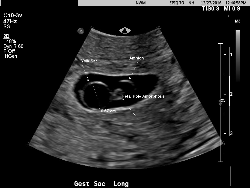
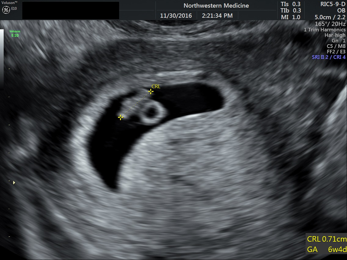
Fetal pole | Radiology Reference Article | Radiopaedia.org